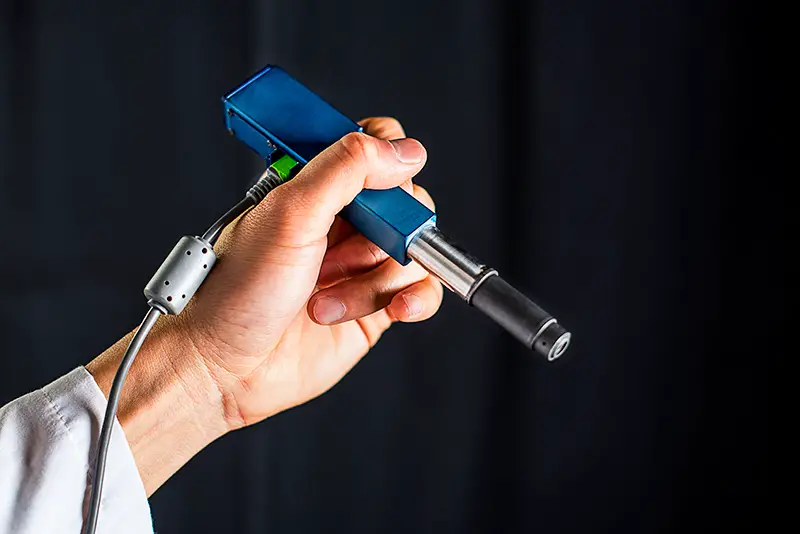
29th January 2016 Pen-sized microscope identifies cancer cells Researchers at the University of Washington have developed a new handheld, pen-sized microscope that could identify cancer cells in doctor's offices and operating rooms.
Surgeons removing a malignant brain tumour don't want to leave cancerous material behind. But they're also trying to protect healthy brain matter and minimise neurological harm. Once they open up a patient's skull, there's no time to send tissue samples to a pathology lab – where they are typically frozen, sliced, stained, mounted on slides and investigated under a bulky microscope – to clearly distinguish between cancerous and normal brain cells. But a handheld, miniature microscope being developed by University of Washington (UW) engineers could allow surgeons to "see" at a cellular level in the operating room and determine precisely where to stop cutting. This technology, made in collaboration with Memorial Sloan Kettering Cancer Centre, Stanford University and the Barrow Neurological Institute, is outlined in the February 2016 issue of Biomedical Optics Express. "Surgeons don't have a very good way of knowing when they're done cutting out a tumour," said Jonathan Liu, senior author on the paper and a UW assistant professor of mechanical engineering. "They're using their sense of sight, sense of touch, pre-operative images of the brain – and oftentimes it's pretty subjective. Being able to zoom and see at the cellular level during the surgery would really help them to accurately differentiate between tumour and normal tissues and improve patient outcomes." Similarly, dentists who find a suspicious-looking lesion in a patient's mouth will often have to cut it out and send it to a lab to be biopsied for oral cancer, a process that subjects patients to an invasive procedure and overburdens pathology labs. A miniature microscope with high enough resolution to see changes at a cellular level could be used in dental or dermatological clinics to assess which lesions or moles are normal and which need to be biopsied.
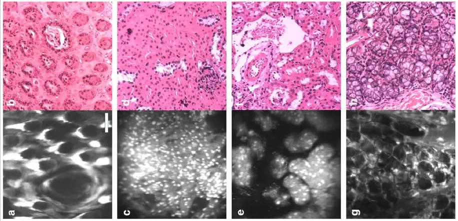
"The microscope technologies that have been developed over the last couple of decades are expensive and still pretty large," said Milind Rajadhyaksha, at the Memorial Sloan Kettering Cancer Centre in NYC, co-author on the study. "So there's a need for creating much more miniaturised microscopes." The new microscope revealed by UW is able to combine technologies in a compact and novel way, generating high-quality images at faster speeds than existing bulkier devices. It uses "dual-axis confocal microscopy" to illuminate and more clearly see through opaque tissue, capturing details up to half a millimetre beneath the tissue surface, where some types of cancerous cells originate. In the video below, for example, the team produced images of fluorescent blood vessels in a mouse ear. In their paper, they demonstrate how their invention has sufficient resolution to see subcellular details. "For brain tumour surgery, there are often cells left behind that are invisible to the neurosurgeon. This device will really be the first to let you identify these cells during the operation and determine exactly how much further you can reduce this residual," said project collaborator Nader Sanai, a professor of neurosurgery at the Barrow Neurological Institute in Phoenix. "That's not possible to do today." Human clinical trials are expected to start in 2017 and the team hopes it can be introduced into surgeries by 2018-2020.
Comments »
|