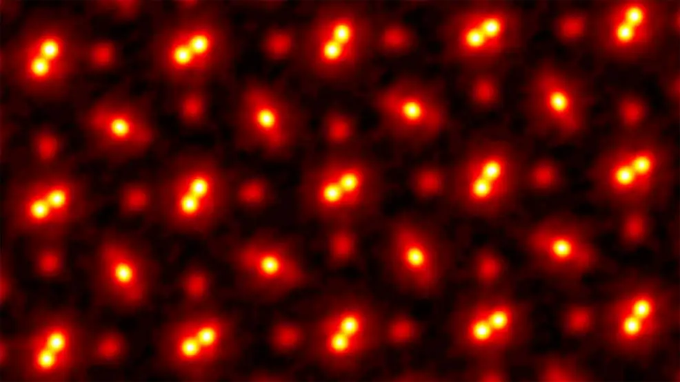
23rd May 2021 Atoms viewed at highest ever resolution Researchers at Cornell University have developed a new microscopy technique that can shrink spatial resolutions to less than 20 picometres.
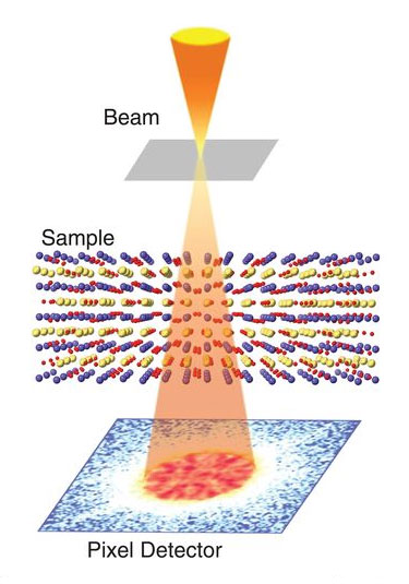
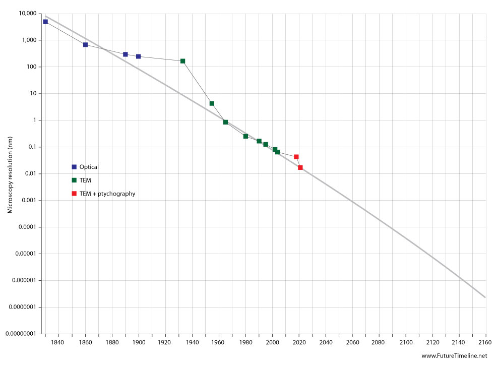
Transmission electron microscopy (TEM) is a technique that involves beaming electrons through a specimen to form an image. This enables the generation of significantly higher resolution than traditional optical microscopes. While the latter devices are typically limited to around 1000x magnification due to the resolving power of visible light, TEM can provide zoom capabilities that are orders of magnitude greater – surpassing even a scanning electron microscope (SEM). In recent years, TEM instruments have begun to reach extraordinary levels of detail. Spatial resolutions are now edging into the realm of individual atoms, measuring less than 0.0000005 millimetres (mm). However, TEM is prone to lens aberrations and multiple scattering, limiting its use to samples thin enough to let electrons pass through. The process is technically challenging and requires additional tools to perform. In 2018, researchers at Cornell University offered a potential solution. They built a high-powered detector combined with a new algorithm-driven process called ptychography. This achieved a new record for microscopic resolution, tripling the previous state-of-the-art. Now the same team has done it again, shrinking the spatial resolution down from 50 picometres (pm) to less than 20 pm. This time, the researchers developed an electron microscope pixel array detector (EMPAD) that incorporates even more sophisticated 3D reconstruction algorithms. The imaging is so fine-tuned that, according to the researchers, the only blurring that remains is caused by thermal fluctuations in the atoms themselves.
"This doesn't just set a new record," said David Muller, Professor of Engineering at Cornell. "It's reached a regime which is effectively going to be an ultimate limit for resolution. We basically can now figure out where the atoms are in a very easy way. This opens up a whole lot of new measurement possibilities of things we've wanted to do for a very long time. It also solves a long-standing problem – undoing the multiple scattering of the beam in the sample, which Hans Bethe laid out in 1928 – that has blocked us from doing this in the past." Ptychography works by scanning overlapping scattering patterns from a material sample and looking for changes in the overlapping region. The team have described their method, used on a crystal of praseodymium orthoscandate (PrScO3), in a paper that appears this week in the journal Science. "We're chasing speckle patterns that look a lot like those laser-pointer patterns that cats are equally fascinated by," explained Muller. "By observing how the pattern changes, we are able to compute the shape of the object that caused the pattern. "With these new algorithms, we're now able to correct for all the blurring of our microscope to the point that the largest blurring factor we have left is the fact that the atoms themselves are wobbling, because that's what happens to atoms at finite temperature. When we talk about temperature, what we're actually measuring is the average speed of how much the atoms are jiggling." The team believes their record may be topped again by using heavier atoms, which wobble less, or by cooling down the sample. Even at zero temperature, however, atoms still have quantum fluctuations. Although their method is time-consuming and computationally demanding, it could be made more efficient with more powerful computers in conjunction with machine learning and faster detectors. The new imaging technique will provide numerous applications. It could enable scientists to locate individual atoms in all three dimensions when they might otherwise be hidden using other methods. Researchers will also be able to find "impurity" atoms in unusual configurations and image them and their vibrations, one at a time. The imaging of semiconductors, catalysts and quantum materials will be greatly improved – including those used in quantum computing – as well as imaging of atoms at the boundaries where materials are joined together. Biological cells, tissues, or even the synapse connections in the brain will also be viewable in greater detail. With microscopy having reached the level of atoms, we may have to wait a long time until the next big milestone in this field. Rather like the differences between planetary, Solar System, interstellar, and galactic scales, the inner components of an atom are separated by orders of magnitude. Going by the current trend in spatial resolution, reaching the scale of protons could take until the mid-22nd century. We would have to wait even longer for the scale of quarks and electrons, which are three orders of magnitude smaller than protons and a whopping eight orders of magnitude smaller than a complete atom.
Comments »
If you enjoyed this article, please consider sharing it:
|