16th April 2016 Artificial intelligence finds cancer cells more efficiently By using a laser at nanosecond speeds, in combination with deep learning algorithms, a new microscope detects cancer cells more efficiently than standard methods.
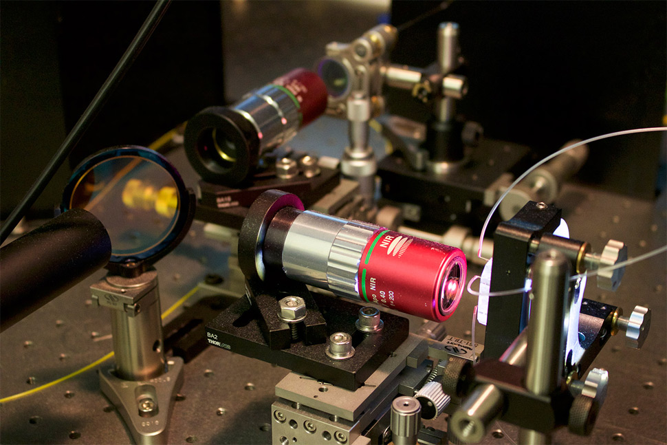
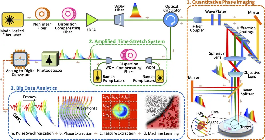
Scientists at the University of California, Los Angeles (UCLA) have developed a new technique for identifying cancer cells in blood samples, faster and more accurately than current standard methods. One common approach to testing for cancer involves doctors adding biochemicals to blood samples. These biochemicals attach biological "labels" to cancer cells, which enable instruments to detect and identify them. However, the biochemicals can damage cells and render the samples unusable for future analyses. Other techniques are available that don't use labelling, but these can be inaccurate, because they only identify cancer cells based on a single physical characteristic. The new technique, demonstrated by the California NanoSystems Institute at UCLA, images cells without destroying them. Not only that, but it can identify up to 16 physical characteristics – including size, granularity and biomass – instead of just one. It combines two components that were invented at UCLA: a photonic time stretch microscope, for rapidly imaging cells in blood samples, and a deep learning program that identifies cancer cells with over 95 percent accuracy. The "photonic time stretch" was invented by Professor Barham Jalali, who holds a patent for this technology, and its use in microscopes is just one of many possible applications. It works by taking pictures of flowing blood cells using laser bursts in the way that a camera uses a flash. This process happens so quickly – in nanoseconds, or billionths of a second – that the images would be too weak to be detected and too fast to be digitised by normal instrumentation. The new microscope overcomes those challenges using specially designed optics that boost the clarity of the images and simultaneously slow them enough to be detected and digitised at a rate of 36 million images per second. It then uses deep learning to distinguish the cancer cells from healthy white blood cells. Deep learning is a form of artificial intelligence that uses complex algorithms to extract meaning from data, with the goal of achieving accurate decision making.
"Each frame is slowed down in time and optically amplified so it can be digitised," explains Ata Mahjoubfar, a UCLA postdoctoral fellow. "This lets us perform fast cell imaging that the artificial intelligence component can distinguish." Normally, taking pictures in such minuscule periods of time would require intense illumination, which could destroy live cells. The UCLA method eliminates that problem too: "The photonic time stretch technique allows us to identify rogue cells in a short time with low-level illumination," said Claire Lifan Chen, a UCLA doctoral student. In their paper – published in the journal Nature Scientific Reports – the researchers write that their system could lead to data-driven diagnoses by the cells’ physical characteristics, which could allow quicker and earlier diagnoses of cancer, for example, and a better understanding of the tumour-specific gene expression in cells, leading to new treatments for disease. ---
Comments »
|